Diagnostic Imaging Center
Advantages of MRI scans in High Definition at Texas Neurology
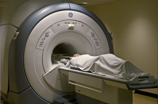
- Our 3.0T and 1.5T High Definition (HD) MRI scanners from General Electric provide the highest clinical field strength and resolution available in the metroplex.
- Our protocols are tailored to patient diagnosis or symptoms.
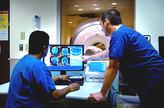
- Our board-certified radiologists from American Radiology Associates with subspecialties in neurology, musculoskeletal, orthopedic, vascular and cardiac disorders interpret all studies.
- We offer the convenience of same day service for patients being referred for both a neurological consultation and an MRI scan.
(includes radiological interpretation)
(includes radiological interpretation)
- In-Network
We are in-network with all major insurance carriers. - Caring and Helpful Staff
Our staff are highly trained individuals in their fields of expertise. - Comfortable
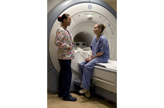
Our scanners provide increased comfort and easier exam positions; wheelchair accessible. - Efficient
Our scanners reduced the need to rescan or perform redundant exams due to patient or organ motion.
- Hassle-Free Scheduling
Fax your order along with patient demographics to (214) 827-4343 and our referral staff will handle scheduling, obtain pre-authorization and complete the appropriate patient safety screening. - Results Report
Results are provided within 24 hours via fax or email. CD images or films as well as online access to images and reports can be provided upon request. - STAT Requests
MRI results can be provided by phone in a matter of hours.

-
Self-parking is available on the 4th level in the parking garage above our building in the spots with a blue wheel stop with white "NEURO PATIENT" letters.
Utilize the elevator on the north side of the building. After exiting, proceed through the door on the left into our main lobby where you'll be greeted by a staff member and receive additional guidance on checking in.
A short-term patient drop off lane is available in the front of the building for patients with mobility issues. -
Phone
-
Fax(214) 827-4343
-
Mon7:30am to 7:30pmTue7:30am to 7:30pmWed7:30am to 7:30pmThu7:30am to 7:30pmFri7:30am to 7:30pmSat7:30am to 4:30pmSunClosed
Overview
Introduction
The human musculoskeletal system consists of the human skeleton, made by bones attached to other bones with joints, and skeletal muscle attached to the skeleton by tendons.
Background
Some musculoskeletal disorders affect primarily the joints, causing arthritis. Others affect primarily the bones (eg, fractures, Paget's disease, tumors), muscles or other extra-articular soft tissues (eg, polymyalgia rheumatica, fibromyalgia), or periarticular soft tissues (eg, bursitis, tendinitis, sprain). Arthritis has myriad possible causes, including infection, autoimmune disease, crystal-induced inflammation, other kinds of inflammation, and noninflammatory tissue degeneration; systemic diseases may be involved. Arthritis may affect single or multiple joints, in either a symmetric or asymmetric manner.
Pain is the most common symptom of many joint disorders. Usually, pain originating from superficial structures is better localized than pain originating from deeper structures. Pain originating in small distal joints tends to be better localized than pain originating in large proximal joints. Joint pain can be referred from extra-articular structures or from other joints. Arthritis often produces "aching" pain, whereas neuropathies often produce "burning" pain.
"Stiffness" may mean weakness, fatigue, or fixed limitation of motion to patients. Stiffness of importance to rheumatic disease is difficulty in motion that develops when attempting to move a joint after a period of rest. The duration of stiffness after beginning joint motion reflects its severity. Stiffness is more severe in inflammatory joint disorders. Morning stiffness can be an important early symptom of RA.
Fatigue is a desire to rest that reflects exhaustion. It differs from weakness, inability to move, and reluctance to move due to pain with movement.
Instability, or buckling of a joint, may suggest weakness of the ligaments or other structures that stabilize the joint, which are assessed by stress testing. Buckling occurs most often in the knee and most often results from an internal joint derangement.
MKS Imaging
For musculoskeletal imaging, plain x-rays may be obtained first, but these are often less accurate than CT or MRI. MRI is the most accurate study for fractures not visible on plain x-rays, particularly in the hip and pelvis, and for soft tissues and internal derangements of the knee.
FAQ
MRI allows doctors to see images of your internal organs and structures in great detail from many angles. This gives them information more quickly, and in many cases more economically, than past tests and exploratory surgeries.
No. Since MRI is "non-invasive", the exam is painless. However, your doctor may utilize a contrast agent to better visualize a part of your anatomy. If this is the case, you may receive a simple shot prior to or during the exam.
No, but you will hear a loud knocking or buzzing sound at various intervals throughout your exam. Other than that, you won't feel a thing.
No. MRI uses a powerful magnet in conjunction with radiofrequency waves to generate images of your internal organs and structures. There is no ionizing (X-Ray) radiation.
There are very few patients who cannot be comfortably accommodated for an MRI exam.
Yes. All MR systems are open at both ends but some also have wider openings on the sides.
That will depend on your height and what part of your body is being scanned. The part that is being imaged is in the middle of the magnet. For example, if your ankle is being scanned, your head will be outside of the MR scanner. If it is your head, neck, or chest is scanned, your head will be inside of the scanner.
Yes. Our MRI system uses a special mirror arrangement to allow you to see outside the magnet at all times.
MRI procedures usually take 45 minutes, but may require additional time, depending on the area scanned.
In most cases, you can just stick with your normal, everyday routine - no special preparation is needed. You can eat and drink your usual diet, work, or play sports (unless you have an injury!) - and take any prescription medications you need. However, there may be some circumstances in which you'll be given specific instructions to follow before the exam. These will be given to you by your doctor, or assistant.
Yes. Because the MRI machine uses a strong magnetic field, which will move objects made with iron or steel, let your doctor and the MRI technologist know if you have:
- A pacemaker
- Aneurysm clips
- Cochlear implants
- A neuro-stimulator (Tens-unit)
- Metal implants
- Steel surgical staples or clips
- An implanted drug infusion device
- Any implant made partially or wholly of iron or steel
- Also, if you're pregnant, let the doctor know.
Even metal objects made of iron or steel can interfere with the exam - so don't bring any of the following into the examination room (a secure place to store your valuables will be provided):
- Coins
- Jewelry
- Watches
- Keys
- Dentures or partial plates
- Hearing aids
Magnetic waves can also erase the code on bank cards and credit cards, so don't bring any to your exam. Last of all, you will be asked to change into a patient gown.
Most people have no reaction at all. However, if you have had claustrophobic reactions to enclosed spaces before, you should let the doctor or technologist know. Even if you are uncomfortable in small spaces, staff members can help you complete the study.
You will be in contact with a technologist at all times. Even when he or she is not in the MRI room, you will be able to talk to him or her by intercom. The technologist is always able to see you through a large patient viewing window. In some cases a friend or family member may stay in the scan room with you during your exam. Please consult the Diagnostic Imaging Center staff if you wish to have someone accompany you during the exam.
The magnet makes a knocking sound as images are being taken. In between scans the machine is quiet. Ear plugs will provided available to you for your exam and their use will not prevent you from hearing the technologist if he or she speaks to you during the exam.
You do have to remain as still as possible, but the time passes quickly. Moving during the procedure may require repeating parts of the exam so it is best to try to remain as still as possible for the best exam results.
That will depend on what is being studied, but a typical exam lasts between 30 and 60 minutes. You should always allow extra time in case the exam lasts longer than expected.
Most insurance plans will reimburse the cost of most MRI exams. To find out if your insurance plan covers the specific MRI exam you will be having, contact your insurance carrier. MRI exams are frequently subject to deductibles and co-insurance which are a separate benefit from your physician's services.
Some patients with metal implants cannot be safely scanned in the MR environment. People with pacemakers, aneurysm clips, especially in the brain; and neurostimulators generally cannot be scanned. Anyone with surgical pins, shrapnel, plates or other type of metal implants should notify the technologist. You will be required to provide a health history when you arrive for your exam explaining any metallic implants you may have. A doctor will determine if a particular metal implant is approved to be in an MR environment.
To begin the exam, you will lie down on the scan table. When the machine starts to work, you'll hear some loud knocking sounds. These sounds occur whenever the MRI pictures are being taken. Think of them as the clicks a VERY large camera would make when taking pictures! We provide earplugs and headphones to help block out the knocking sounds.
In any case, although it's noisy, an MRI exam is completely painless. The only thing you must do is HOLD STILL. When you take a picture with a camera, your subject must keep still or the picture will come out blurry. It's the same with an MRI machine. If you move, the scans will be out of focus - and you may have to repeat the exam.
If necessary, you may be injected with a solution called a contrast agent. This allows the radiologist to see the image more clearly. MRI contrast agents typically have few or no side effects, and the injection likely will just feel like a slight pinch. You may be asked to give your consent to this injection, at which time a more detailed explanation about the contrast agent will be given to you by the technologist.
Well, you may feel very well rested since you've just been lying on a table and doing absolutely nothing! (In fact, some people even fall asleep during the exam.) Other than that, you'll feel perfectly normal and can go back to your everyday activities. If you have further questions about your MRI exam, the MRI technologist or your doctor will be glad to answer them.
MRI Information
Magnetic resonance imaging (MRI) is a test that produces very clear pictures, or images, of the human body without the use of x-rays. MRI uses a large magnet, radio waves and a computer to produce these images. Your physician may also order an MRA (Magnetic Resonance Angiography). This exam is used to evaluate vascular abnormalities, such as vascular malformations, aneurysms, vertebrovascular and carotid atherosclerosis, thrombosis and dissection. It also lets your doctor see internal organs, blood vessels, muscles, joints, tumors, areas of infection, and more without x-rays, surgery, or pain. MRI is very safe; in fact, it makes use of natural forces and has no known harmful effects. It's important to know that an MRI will not expose you to any radiation.
What is an MRI?
Magnetic Resonance Imaging (MRI) is a diagnostic imaging technology that uses a strong magnet and radiofrequency waves to produce pictures or "images" of your internal organs and structures. Because MRI allows your doctor to see inside your body from any angle with great clarity, it is giving doctors a wealth of information more quickly and in many cases, more economically than past tests and exploratory surgeries.
Magnets and Metal Don't Mix
When you first enter the MR clinic, you must let the staff and technologist know if you have a pacemaker, surgical clips, prosthesis, metal implants or any other metal objects in your body. Some implants (e.g., a pacemaker) may be affected by an MR examination. The clinic personnel will then determine whether or not you should proceed with the MR examination.
Any metal materials that might be affected or attracted by the powerful magnet used for MR imaging should be left at home or can be secured in a locked cabinet in your private changing area. This list includes your watch, coins, keys, bobby pins, credit cards, pocketknives, etc. You should also be certain that you are reasonably clean of metal flakes or slivers on your skin, as found in some eye make-up or as a result of working around metal finishing or grinding equipment.
Your MRI Exam
Your MR exam is really quite simple. With the assistance of an MR technologist, you will be positioned on a padded table. Because it is essential that you remain as still as possible. The padded table will move smoothly into the magnet opening and your exam will begin. The technician is able to speak with you at all times during the exam.
During your MRI exam you won't feel anything. The only thing you'll notice is a knocking or buzzing sound that occurs as the images are being taken. You will be provided with earplugs to wear during the scan, or an MR compatible audio headset may be available to minimize the noise. The length of your exam is dependent on the type of study being done but will generally last from 45 minutes to over an hour.
An intercom and mirror arrangement in the system allows the MR staff to see and hear you throughout the exam. If you become uncomfortable at any point, you can alert the technician. The MR staff will be right there to assist you. Once the exam is completed, your technologist will bring you back to the preparation room to collect your belongings. And that's all there is to it!
For more information on Diagnostic Imaging, download our "What You Need to Know about Your MRI" brochure.
Testing Procedures
An MRI examination poses no risk to the average patient, if appropriate safety guidelines are followed. Patients who have the following conditions can be safely examined with an MRI:
- Surgical clips or sutures
- Post-cardiac surgical patients
- Artificial joints
- Staples
- Cardiac valve replacements (except the Starr-Edwards metallic ball/cage)
- Disconnected medication pumps
- Vena cava filters
- Brain shunt tubes for hydrocephalus
Personal items such as your watch, wallet, including any credit cards with magnetic strips (they will be erased by the magnet), and jewelry should be left at home if possible, or removed prior to the MRI scan. Secured lockers are available to store personal possessions. Allow 2 hours for your MRI exam. In most cases, the procedure takes 40 to 80 minutes, during which several dozen images may be obtained.
You will be asked to wear a hospital gown during the MRI scan. As the MRI scan begins, you will hear the equipment making a muffled thumping sound which will last for several minutes. Other than the sound, you should experience no unusual sensations during the scan.
*For certain exams, an injection of a contrast material (OMNISCAN) may be administered. This helps identify certain anatomic structures on the scan images. (*Asymptomatic, transitory changes in serum iron and calcium have been observed in patients who have received OMNISCAN. The clinical significance is unknown. OMNISCAN interferes with serum calcium measurements with some methods commonly used in hospitals, resulting in serum calcium concentrations lower than true values. It is recommended not to use such methods for 12-24 hours after administration of OMNISCAN.)
If you must have laboratory testing 24-48 hours after your MRI, please notify your physician that you have received MRI contrast. Please feel free to bring up any questions or concerns to the technologist or the physician.
Generally, you will be able to resume normal activity immediately after the procedure. The results of the MRI should be available to your physician 5 days after your test is completed, Monday through Friday. Please remember, we are happy to discuss your questions and concerns. Please feel free to ask.
Special Precautions
MRI testing may be inadvisable for patients with certain conditions. Please tell your doctor if you have any of the following conditions:
- A heart pacemaker
- A cerebral aneurysm clip (metal clip on a blood vessel in the brain)
- You are pregnant
- An implanted insulin pump (for treatment of diabetes), an implanted narcotics pump (for pain medication, or implanted nerve stimulators ("TENS") for back pain
- Metal in your eye or eye socket
- A cochlear (ear) implant for hearing impairment
- Implanted spine stabilization rods
- Severe lung disease (such as tracheomalacia or bronchopulmonary dysplasia)
- Body weight greater than 350 pounds
- Inability to lie on back for 30 to 60 minutes
- Claustrophobia (fear of closed or narrow spaces)
Documents & Forms
- Medical Appointment Cancellation & No Show Policy
- 52 Proven Stress Reducers
- Authorization to Disclose Protected Health Information Form (To Obtain) (Dallas Version)
- Authorization to Disclose Protected Health Information Form (To Release) (Dallas Version)
- Chronic Pain or Illness: Relationships and Communication
- Commonly Reported Symptoms at Various Phases of Migraine and Chronic Daily Headache
- The Complete Headache Chart
- Distinguishing Sinus Headache From Migraine
- Emergency Room Treatment Form
- Financial Policy
- Food Additives And Migraines
- General Guidelines for Treatment with Preventative Medications
- Headache Calendar
- Infusion Order Form (Ocrevus (Ocrelizumab))
- Low Tyramine Headache Diet
- Menstrually Related Migraine
- MRI Screening Form
- New Patient Packet (Dallas Version)
- Notice of Privacy Practices (English Version) (Spanish Version)
- Nurse Practitioner/Physician Assistant Information Guide
- Patient Authorization Form for Radicava IV Infusion
- Questionnaire (Intake)
- Referral Form (Diagnostic Imaging Center)
- Referral Form (General)
- The Role of Triggering Factors in Migraine Headaches
- Sinus Headache or Migraine?
- Steps to Take to Reduce the Impact of Migraine at Work
- TeleVisit Terms & Conditions
- Ten Day Prednisone Program
- Tips for Better Sleep
- Tips for Improving the Inner "Landscape" or Increasing Self-Esteem
- Travel Tips for the Headache Sufferer
- Treatment Strategies and Options for Chronic Daily Headache Sufferers
- What Triggers A Migraine
- What You Need to Know about Your MRI
- Oops! It doesn't look like there's anything here right now...
- Headache Relief for Women by Dr. Alan Rappaport
- Heal Your Headache by Dr. David Buchholtz
- The Migraine Brain by Dr. Carolyn Bernstein
- Tell Me What to Eat/Headache by Elaine McGee R.D.
Resources
- Alzheimers Foundation
- American Academy of Neurology
- American Academy of Sleep Medicine
- American Council for Headache Education (ACHE)
- American Headache and Migraine Association (AHMA)
- American Medical Association
- American Parkinson Disease Association
- American Stroke Association
- Beeman Hotel Reservation Link
- BOTOX Chronic Migraine
- CenterWatch
- Clinical Trials Sites
- The Consortium of Multiple Sclerosis Centers (CMSC)
- Dystonia Medical Research Foundation
- Eunice Kennedy Shriver National Institute of Child Health and Human Development
- Everyday Life with ALS book
- The Foundation for Peripheral Neuropathy
- The Impact of Migraine: Q&A with Dr. David Dodick
- International Organization of Multiple Sclerosis Nurses (IOMSN)
- Medicare
- The Movement Disorder Society
- The Multiple Sclerosis Association of America (MSAA)
- Multiple Sclerosis International Foundation
- Muscular Dystrophy Association (MDA) - ALS Division
- MyChronicMigraine
- National ALS Registry
- National Center for Complementary and Alternative Medicine
- National Institute for Neurological Disorders and Stroke (NINDS)
- National Institute of Neurological Disorders and Stroke (NINDS) (ALS Information Page)
- Northeast ALS Consortium (NEALS)
- Parkinson Disease Foundation
- Quack Watch
- Washington University
- We Move
- Oops! It doesn't look like there's anything here right now...